
This manual serves as a comprehensive guide, initiating your exploration of human anatomy and physiology through practical laboratory experiences and detailed instructions.
Burns’ 2E edition, published in 2009, provides essential resources for understanding physiological concepts and anatomical structures within a laboratory setting.
Begin your journey with this manual, designed to facilitate successful operation and enjoyment while mastering the intricacies of the human body.
Purpose of the Lab Manual
The primary purpose of this Human Anatomy & Physiology Laboratory Manual is to complement and enhance your understanding of the core concepts presented in the lecture portion of the course.
Through hands-on activities, dissections, and microscopic observations, you will gain practical experience applying theoretical knowledge to real-world anatomical structures and physiological processes.
This manual guides you through each experiment, providing clear instructions, detailed illustrations, and critical thinking questions to foster a deeper comprehension of the human body’s intricate workings.
Ultimately, it aims to develop your laboratory skills, analytical abilities, and appreciation for the remarkable complexity of human anatomy and physiology.
Scope of Coverage
This lab manual comprehensively covers a wide range of topics within human anatomy and physiology, mirroring the foundational elements of a typical introductory course.
It encompasses all major body systems, including the integumentary, skeletal, muscular, nervous, endocrine, cardiovascular, respiratory, digestive, urinary, and reproductive systems.
The manual delves into histological examination of tissues, anatomical dissections, and physiological experiments designed to illustrate key functional principles.
Burns’ 2E edition, published in 2009, provides a robust framework for exploring human structure and function through practical laboratory investigations.
Safety Precautions in the Lab
Prioritizing safety is paramount within the anatomy and physiology laboratory environment. Always adhere to all instructor guidelines and established protocols.
Proper handling of specimens, chemicals, and dissection tools is crucial to prevent injury and contamination. Wear appropriate personal protective equipment (PPE), including gloves and eye protection.
Dispose of biological waste correctly, following designated procedures. Report any accidents or spills immediately to the instructor. Maintain a clean and organized workspace.
Understanding and implementing these precautions ensures a safe and productive learning experience for everyone.

II. Basic Anatomical Terminology
Mastering anatomical language is fundamental for precise communication regarding body structures and their relationships, essential for lab work and understanding.
Anatomical Position & Planes
Understanding the standard anatomical position – standing erect, feet slightly apart, palms facing forward – is crucial for consistent descriptions of body parts and directions.
Anatomical planes divide the body, enabling visualization of internal structures. Sagittal planes separate left and right, frontal planes divide anterior and posterior, and transverse planes create superior and inferior sections.
These planes are fundamental for interpreting imaging, performing dissections, and accurately communicating anatomical relationships within the laboratory setting, ensuring clarity and precision in your studies.
Directional Terms (Superior, Inferior, etc.)
Precise anatomical language utilizes directional terms to describe body part locations relative to one another. Superior indicates above, while inferior denotes below. Anterior refers to the front, and posterior to the back.
Medial signifies near the midline, and lateral means farther from it. Proximal and distal describe closeness to or distance from a limb’s origin.
Mastering these terms, as outlined in the lab manual, is essential for accurate communication and understanding of anatomical relationships during dissections and studies.
Body Cavities (Dorsal & Ventral)
The human body contains cavities protecting delicate organs. Dorsal cavities include the cranial (brain) and vertebral (spinal cord) – providing bony protection. Ventral cavities are larger and include the thoracic and abdominopelvic.
The thoracic cavity houses the heart and lungs, while the abdominopelvic cavity contains abdominal organs and pelvic structures.
Understanding these divisions, detailed within the lab manual, is crucial for visualizing organ placement and appreciating anatomical relationships during dissection exercises.

III. Histology: The Study of Tissues
This section explores tissues – groups of similar cells performing specific functions. The lab manual guides microscopic observation and identification of tissue types.
Epithelial Tissue Types & Functions
Epithelial tissues, covering body surfaces and lining cavities, are a primary focus of histological study within this lab manual. Students will learn to differentiate between various types – squamous, cuboidal, and columnar – based on cell shape and arrangement.
The manual details how these tissues perform crucial functions like protection, absorption, secretion, and excretion. Microscopic examination of prepared slides will reveal structural characteristics, aiding in identification. Understanding simple versus stratified arrangements, and the presence of cilia or microvilli, is emphasized for functional correlation.
Practical exercises will reinforce the link between epithelial structure and its specific role within the body.
Connective Tissue Types & Functions
This lab manual extensively covers connective tissues – the most abundant tissue type – focusing on their diverse structures and critical roles. Students will identify connective tissue proper (loose and dense), supporting connective tissues (cartilage and bone), and fluid connective tissues (blood).
Emphasis is placed on recognizing the extracellular matrix, composed of ground substance and fibers, and how it dictates tissue properties. Microscopic observation of prepared slides will highlight variations in cell types and fiber density. Functions like binding, support, protection, and transport are thoroughly explored.
Practical exercises solidify understanding through identification and functional correlation.
Muscle Tissue Types & Functions
The lab manual details the three primary muscle tissue types: skeletal, smooth, and cardiac, emphasizing their unique structural and functional characteristics. Students will examine microscopic slides to differentiate these tissues based on cell shape, striations, and control mechanisms.
Practical exercises focus on identifying muscle fibers and understanding their roles in movement, posture, and internal processes. Key concepts include excitation-contraction coupling and the sliding filament theory. Functions such as voluntary control, involuntary contractions, and rhythmic pumping are explored.
Detailed observations enhance comprehension.
Nervous Tissue: Neurons & Glial Cells
This section of the lab manual focuses on the fundamental components of the nervous system: neurons and glial cells. Students will learn to identify neuron structures – dendrites, cell body, axon – and understand their roles in signal transmission. Microscopic examination of nervous tissue reveals diverse neuronal morphologies.
Glial cells, including astrocytes, oligodendrocytes, and microglia, are explored for their supportive and protective functions. Practical exercises emphasize the importance of these cells in maintaining neuronal health and facilitating synaptic communication. Understanding their interplay is crucial.

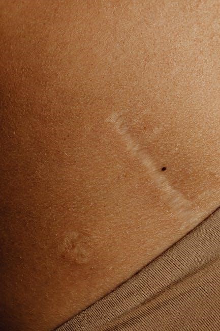
IV. The Integumentary System
This lab manual section details the skin’s layers – epidermis, dermis, and hypodermis – alongside accessory structures like hair, nails, and glands for comprehensive study.
Skin Layers (Epidermis, Dermis, Hypodermis)
The epidermis, the outermost layer, provides a protective barrier composed of stratified squamous epithelium, constantly renewed through cell division. Beneath it lies the dermis, a thicker layer containing connective tissue, blood vessels, nerves, and accessory structures like hair follicles and glands.
The dermis supports the epidermis and provides nourishment, while also playing a crucial role in temperature regulation and sensation. Finally, the hypodermis, or subcutaneous layer, anchors the skin to underlying tissues, providing insulation and cushioning with adipose tissue. Lab exercises will focus on identifying these layers microscopically and understanding their unique functions within the integumentary system.
Accessory Structures (Hair, Nails, Glands)
Hair follicles, embedded within the dermis, produce hair shafts composed of keratin, contributing to insulation and protection. Nails, also keratinized structures, protect the distal phalanges and aid in manipulation. Several gland types are integral to skin function:
Sebaceous glands secrete sebum, lubricating the skin and hair. Sudoriferous glands, including eccrine and apocrine glands, produce sweat for thermoregulation and waste excretion. Lab activities will involve identifying these structures histologically and exploring their roles in maintaining skin homeostasis and overall body function.
Functions of the Integumentary System
The integumentary system performs vital functions, including protection against pathogens and UV radiation, temperature regulation through sweat and blood vessel dilation/constriction, and sensory reception via specialized receptors. Vitamin D synthesis occurs upon sun exposure, crucial for calcium absorption.
Lab exercises will demonstrate how the skin acts as a barrier, regulates body temperature, and provides sensory input. Understanding these functions is essential for comprehending overall physiological homeostasis and the skin’s role in maintaining health.
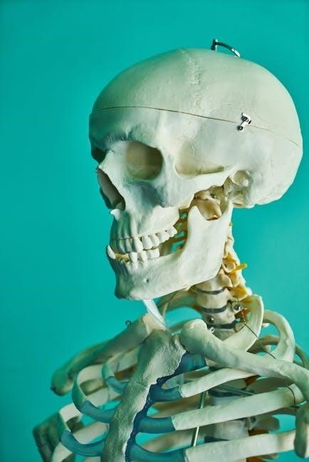
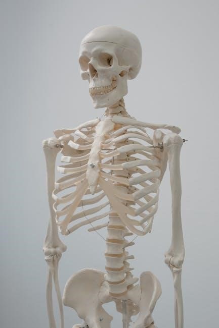
V. The Skeletal System
This section explores bone tissue, structure, classification, and development through lab activities, focusing on the axial and appendicular skeletons for comprehensive understanding.
Bone Tissue & Structure
Bone tissue, a specialized connective tissue, forms the rigid framework of the skeletal system. This lab section delves into its microscopic and macroscopic organization, examining compact and spongy bone structures.
Osteons, the functional units of compact bone, and trabeculae, found in spongy bone, will be explored through microscopic observation. Understanding the composition – including collagen fibers and mineral salts – is crucial.
Labs will focus on identifying bone cells (osteoblasts, osteocytes, osteoclasts) and analyzing bone matrix. Detailed examination of bone structure provides insight into its strength, flexibility, and role in support and protection.
Bone Classification & Development
This lab explores bone classification – long, short, flat, irregular, and sesamoid – relating structure to function. Students will identify examples of each type and analyze their unique characteristics.
Ossification, the bone development process, is a key focus, covering both intramembranous and endochondral ossification. Microscopic slides will illustrate cartilage templates and bone matrix formation.
Labs will demonstrate how bone growth plates facilitate longitudinal growth and how remodeling maintains bone integrity. Understanding these processes is vital for comprehending skeletal development and adaptation throughout life.
Axial Skeleton (Skull, Vertebral Column, Rib Cage)
This lab focuses on the axial skeleton’s components: the skull, vertebral column, and rib cage, providing crucial body support and protection. Students will identify cranial and facial bones, sutures, and foramina.
Vertebral column dissection will highlight regional variations – cervical, thoracic, lumbar, sacral, and coccygeal – and associated ligaments. Rib cage analysis will cover true, false, and floating ribs.
Labs emphasize articulating these structures and understanding their functional relationships, crucial for movement and organ protection. Detailed models and diagrams will aid in anatomical identification;
Appendicular Skeleton (Limbs & Girdles)
This lab explores the appendicular skeleton – limbs and girdles – responsible for movement and manipulation. Students will identify bones of the pectoral and pelvic girdles, upper and lower limbs.
Focus will be on bone markings, articulating surfaces, and muscle attachment sites. Dissection will highlight the structural relationships between bones, ligaments, and joints.
Labs emphasize understanding limb movements and the functional significance of girdle attachments. Detailed models and comparative anatomy exercises will enhance comprehension of skeletal diversity.
VI. The Muscular System
This section details muscle tissue types – skeletal, smooth, and cardiac – alongside contraction mechanisms and major skeletal muscle actions for practical application.
Muscle Tissue Types (Skeletal, Smooth, Cardiac)
Skeletal muscle, attached to bones, enables voluntary movements through striated fibers and controlled contractions, crucial for locomotion and posture.
Smooth muscle, found in organ walls, facilitates involuntary functions like digestion and blood vessel constriction, lacking striations for sustained contractions.
Cardiac muscle, exclusive to the heart, exhibits rhythmic, involuntary contractions due to specialized cells and intercalated discs, ensuring continuous blood circulation.
Laboratory exercises will focus on identifying these tissues microscopically, understanding their structural differences, and correlating structure with specific physiological roles within the body.
Muscle Contraction Mechanism
Muscle contraction arises from the sliding filament theory, where actin and myosin filaments interact, shortening the sarcomere—the functional unit of muscle.
Neuromuscular junctions release acetylcholine, initiating action potentials that travel along the muscle fiber, triggering calcium release from the sarcoplasmic reticulum.
Calcium ions bind to troponin, exposing myosin-binding sites on actin, enabling cross-bridge formation and the power stroke, ultimately causing contraction.
Lab activities will demonstrate these processes through models and simulations, enhancing understanding of the intricate steps involved in muscle function.
Major Skeletal Muscles & Their Actions
This section details key skeletal muscles, including the biceps brachii (flexion), quadriceps femoris (extension), and gastrocnemius (plantar flexion), among others.
Lab exercises involve identifying these muscles on anatomical models and cadavers, correlating their structure with specific movements.
Understanding origins, insertions, and actions is crucial for comprehending how muscles produce movement at joints, enabling functional analysis.
The manual provides detailed illustrations and descriptions to aid in muscle identification and action comprehension during laboratory sessions.

VII. The Nervous System
This section explores neuron structure, brain anatomy, and spinal cord function, utilizing lab exercises for comprehensive understanding of nervous system components.
Neuron Structure & Function
This lab focuses on the fundamental units of the nervous system – neurons. Students will identify and analyze key neuronal components, including the cell body (soma), dendrites, axon, and myelin sheath, understanding their roles in signal transmission.
Practical exercises will involve microscopic observation of neuron types and tracing the pathway of nerve impulses. Emphasis is placed on understanding how structure dictates function, exploring concepts like action potentials and synaptic transmission.
The manual guides students through experiments demonstrating neuronal communication, solidifying their grasp of this critical physiological process and its impact on overall nervous system activity.
Brain Anatomy & Function
This section delves into the complex organization of the human brain, utilizing models and diagrams to identify major regions like the cerebrum, cerebellum, and brainstem. Students will explore the lobes of the cerebrum and their associated functions – motor, sensory, and association areas.
Lab activities include tracing neural pathways and correlating brain regions with specific behaviors and cognitive processes. The manual provides detailed illustrations and explanations to aid in comprehension.
Understanding the intricate relationship between brain structure and function is paramount, preparing students for advanced neurological studies.
Spinal Cord Anatomy & Function
This lab focuses on the structural components of the spinal cord, including its protective meningeal layers and the organization of gray and white matter. Students will identify dorsal and ventral horns, and explore the pathways for sensory and motor information.
Activities involve tracing nerve roots and spinal nerves, understanding their connection to specific body regions. The manual provides detailed diagrams illustrating reflex arcs and the transmission of signals.
Emphasis is placed on the spinal cord’s role as a crucial link between the brain and peripheral nervous system.

VIII. The Endocrine System
This section explores hormone types, mechanisms of action, and major endocrine glands, utilizing the lab manual for detailed study and practical application.
Hormone Types & Mechanisms of Action
The lab manual details diverse hormone classifications, including steroid, protein, and amine hormones, each exhibiting unique chemical structures and synthesis pathways.
Mechanisms of action are thoroughly explained, covering receptor binding – both intracellular for steroids and membrane-bound for proteins – initiating signaling cascades.
Explore how hormones regulate physiological processes via positive and negative feedback loops, maintaining homeostasis within the body’s intricate systems.
Practical exercises within the manual will reinforce understanding of hormone interactions and their impact on target tissues, enhancing comprehension of endocrine control.
Major Endocrine Glands & Their Hormones
The lab manual meticulously examines key endocrine glands – pituitary, thyroid, parathyroid, adrenal, pancreas, and gonads – detailing their anatomical locations and hormonal secretions.
Hormonal profiles are presented, outlining the specific hormones produced by each gland, their chemical structures, and their physiological effects on target organs.
Practical labs focus on correlating gland dysfunction with hormonal imbalances, illustrating clinical relevance and diagnostic approaches.
Understand how these glands orchestrate vital functions like metabolism, growth, reproduction, and stress response through precise hormonal regulation.

IX. The Cardiovascular System
This section details heart anatomy, blood vessel structures (arteries, veins, capillaries), and blood composition, utilizing lab exercises for practical understanding.
Heart Anatomy & Physiology
Explore the heart’s intricate chambers – atria and ventricles – and their coordinated function in pumping blood throughout the body. Lab activities will focus on identifying key structures like valves, major vessels (aorta, vena cava), and the myocardium.
Understand the cardiac cycle, including systole and diastole, and how these phases contribute to efficient blood circulation. Physiological investigations will examine heart rate, blood pressure, and the impact of various factors on cardiovascular performance. This manual provides detailed diagrams and exercises to solidify your comprehension.
Blood Vessels (Arteries, Veins, Capillaries)
This section details the structure and function of arteries, veins, and capillaries – the vital network transporting blood. Lab exercises will involve identifying these vessels microscopically, noting differences in their walls and lumen size. Explore how arteries withstand high pressure, while veins facilitate return flow with valves.
Understand capillary beds as sites of exchange, delivering oxygen and nutrients to tissues. The manual guides you through tracing blood flow pathways and analyzing the relationship between vessel structure and physiological roles in circulation.
Blood Composition & Function
This lab explores blood’s components: plasma, red blood cells, white blood cells, and platelets. Microscopic examination will aid in identifying each cell type and understanding their unique roles. Learn how red blood cells transport oxygen, while white blood cells defend against infection.
The manual details plasma’s role in nutrient transport and waste removal, and platelets’ function in clotting. Investigate hematocrit levels and blood typing, gaining practical skills in analyzing blood’s vital functions within the circulatory system.
X. The Respiratory System
This section details the anatomy of the respiratory tract – nose to lungs – and explores the physiological mechanisms governing efficient breathing processes;
Respiratory Anatomy (Nose, Pharynx, Larynx, etc.)
Detailed exploration of the respiratory system’s structures begins with the nose, responsible for filtering and warming incoming air. The pharynx serves as a passageway for both air and food, connecting to the larynx – the voice box – crucial for sound production.
Further down, the trachea branches into bronchi, leading to the lungs. Within the lungs, bronchioles terminate in alveoli, tiny air sacs where gas exchange occurs. This lab manual will guide you through identifying these structures and understanding their interconnected roles in respiration.
Practical exercises will focus on anatomical models and diagrams, enhancing comprehension of the respiratory pathway’s complex organization.
Mechanisms of Breathing
This section delves into the physiological processes driving respiration – inhalation and exhalation. The lab manual explains how the diaphragm and intercostal muscles contract and relax, altering thoracic cavity volume and creating pressure gradients.
Understanding Boyle’s Law is crucial, demonstrating the inverse relationship between pressure and volume. Practical exercises will involve measuring lung volumes using spirometers, simulating breathing mechanics, and analyzing respiratory rates.
You’ll explore how these mechanisms ensure efficient oxygen intake and carbon dioxide removal, vital for cellular function and overall homeostasis.

XI. The Digestive System
This section explores the organs and processes involved in breaking down food, absorption of nutrients, and elimination of waste, as detailed in the manual.
Digestive Organs & Their Functions
The lab manual meticulously details each digestive organ’s role, starting with the mouth and progressing through the esophagus, stomach, small intestine, and large intestine.
It explains how the stomach utilizes muscular contractions and gastric juices for initial breakdown, while the small intestine focuses on nutrient absorption via villi and microvilli.
Furthermore, the manual clarifies the large intestine’s function in water absorption and waste compaction, culminating in elimination; Accessory organs – liver, gallbladder, and pancreas – are also covered, highlighting their contributions to digestion through enzyme and bile production.
Practical exercises within the manual reinforce understanding of organ anatomy and physiological processes.
Digestive Processes (Absorption, Secretion, Motility)
The lab manual thoroughly examines core digestive processes: absorption, secretion, and motility, detailing their interconnectedness for efficient nutrient extraction.
Absorption, primarily occurring in the small intestine, is explained with emphasis on mechanisms like diffusion, facilitated transport, and active transport of nutrients into the bloodstream.
Secretions – enzymes, hormones, and mucus – are detailed, outlining their source and specific roles in breaking down food and protecting the digestive tract.
Motility, encompassing peristalsis and segmentation, is illustrated, demonstrating how these movements propel and mix food for optimal digestion.
XII. The Urinary System
This section of the lab manual details kidney anatomy, urine formation, and the system’s crucial role in waste removal and homeostasis maintenance.
Kidney Anatomy & Function
The lab manual meticulously explores the kidney’s complex structure, from the outer cortex to the inner medulla, highlighting the nephron as the functional unit.
Detailed diagrams and exercises guide students through identifying key anatomical features like the renal capsule, pyramids, and collecting ducts.
Functional aspects covered include glomerular filtration, tubular reabsorption, and secretion, explaining how the kidneys regulate blood volume, pressure, and electrolyte balance.
Practical exercises may involve examining kidney models, histological slides, and analyzing urine samples to understand normal kidney function and potential pathologies.
Urine Formation
The lab manual dissects the three-stage process of urine formation: glomerular filtration, tubular reabsorption, and tubular secretion, providing a clear understanding of each step.
Exercises focus on calculating glomerular filtration rate (GFR) and analyzing the composition of urine under varying physiological conditions.
Students will learn how the kidneys selectively reabsorb essential substances like glucose and amino acids, while excreting waste products and regulating pH.
Practical applications include identifying factors influencing urine volume and understanding the role of hormones in regulating kidney function.